Top 5 Imaging Trends From ECTRIMS 2019
by Pablo Villoslada, MD, PhD.
The world’s largest meeting on Multiple Sclerosis, ECTRIMS 2019, took place in Stockholm, Sweden. This article reviews the major trends in Imaging of this years’ congress.
1. The central vein sign
The central vein sign has been proposed as a novel imaging biomarker to improve the accuracy and speed of MS diagnosis. However, lack of standardization and validation has prevented its full implementation.

At ETCRIMS 2019 the team from the Université de Lausanne (unil.ch) presented a new automatic method for detecting the central vein sign, which would improve the reproducibility of the technique. In addition, the team from Università Degli Studi Firenze (unifi.it) and ULB Neuroscience Institute in Brussels (uni.ulb.ac.be) showed that the central vein sign may distinguish patients based on their severity. Indeed, the team from Fleni in Buenos Aires (fleni.org.ar) presented the SWAN-venule, a new susceptibility-based MRI technic for improving the sensitivity for detecting this sign. Finally, the team from the University of Toronto (utoronto.ca) presented the association between the central vein sign and the risk of cognitive impairment in patients with radiologically isolated syndromes. In summary, new evidence is being summed up to standardize and support the use of the central vein sign for improving the accuracy and accelerating the speed of MS diagnosis, which has implications in order to start disease-modifying therapies at earlier stages and change the disease prognosis consequently.
2. T2 lesion evolution as a biomarker of progressive disease
T2 lesions are dynamic, showing changes along time. Expansion of T2 lesions has been associated with disease activity or disability worsening and was related to the pathological concept of chronic active lesions or smoldering plaques. The team from Universitat de Girona (udg.edu) presented a convolutional neural network for identifying changes in chronic T2 lesions associated with longitudinal disability worsening. In addition, the team from Valencia provided evidence that T1/T2 ratios are associated with future clinical outcomes. The team from Ospedale San Raffaele in Milan (hsr.it) showed that changes in T2 lesions as slow expanding lesions are modulated by disease-modifying drugs such as fingolimod and natalizumab, which may be associated with lower disability worsening. Although we still lack validation regarding the pathological basis of the changes of T2 lesions, the association with disease course suggests they should be related to some of the key pathogenic mechanisms involves in disease progression.
3. Connectomics in MS

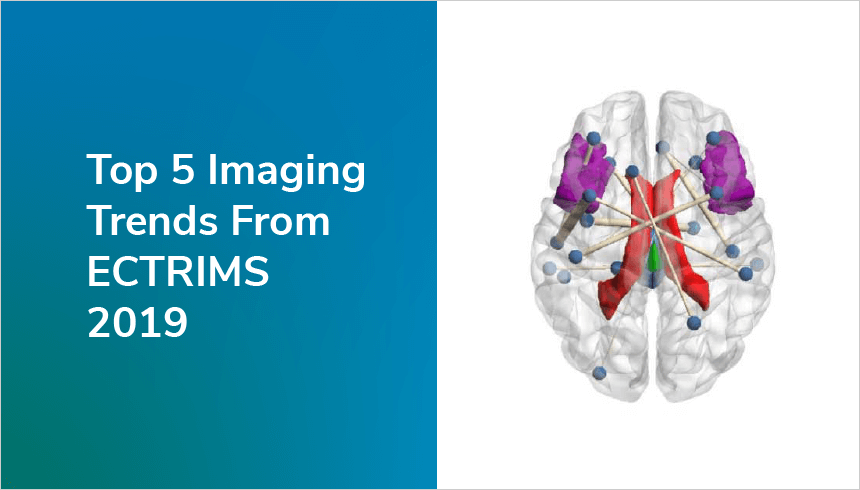
Advances in the Human Connectome Project has provided new tools for interrogating the microstructural damage in the MS brain as well as the preferential damage of specific white matter tracts by the disease. QMENTA (qmenta.com) presented at ECTRIMS his work in collaboration with the team from Barcelona showing that connectomics analysis allows discriminating between healthy and MS brain as well as identifying that long-tracts and commissural tracts are preferentially damaged by the disease. Indeed, statistical models for three relevant brain domains such as walking, vision or cognition were able to explain a significant portion of disability based on tract specific damage. These results were also confirmed in another dataset from the Hospital Clinic of Barcelona (clinicbarcelona.org). Indeed, the team from Boston showed the specific damage of the Medial Longitudinal Fasciculus measured by tractography and the presence of internuclear-ophthalmoplegia, a very robust model of tract-based damage in MS. High-resolution analysis of brain connective and track damage is starting to provide the ability to detect minute damage of the brain and solve the clinical-radiological paradox in MS.
4. Lesion (iron) rim
The presence of iron rims around T2 lesions suggests the presence of activated macrophages phagocyting myelin and for this reason, are considered markers of chronic active plaques
The team from Universität Wien (univie.ac.at)analyzed the presence of iron rims using a 7T magnet in a 7 years longitudinal study, finding an association of iron rim presence with disability accumulation and cognitive impairment. The team from Università di Verona (univr.it) compared two different susceptibility-weighted images protocols for identifying iron rims for improving its identification, whereas the team from Univerzita Karlova in Prague (cuni.cz/uken-1.html) analyze the iron content in deep grey matter as a marker of disease severity. The triad of central vein sign, the T2 slow expanding lesions and iron rims are becoming specific biomarkers for MS diagnosis and prognosis.
5. Imaging demyelination
With the advent of remyelinating drugs, it is critical having imaging markers of demyelination and remyelination. The team from Vrije Universiteit Amsterdam (vu.nl/en) and Cleveland Clinic (my.clevelandclinic.org) analyzed the correspondence between myelin sequences by MRI and the post-mortem pathology in order to validate the substrates of the proposed MRI markers of demyelination. Indeed, the team from Cleveland proposed a multi-modal approach for identifying active demyelination in order to achieve higher accuracy. The team from Boston Massachusetts General Hospital (massgeneral.org) analyzed the presence of cortical demyelination at 7T. Although at present the accuracy of these techniques are still not good enough for monitoring de- and remyelination, new advances are bringing us closer to this aim.
Pablo is a neurologist with more than 20 years of experience in Translational Medicine on biomarkers and new therapeutics for neurological diseases..png?width=2105&height=655&name=catalog-banner%20(1).png)

